Field: Technology
Folate’s Molecular Conduit: Unraveling the Enzymatic Link Between Folic Acid, Vitamin A Signaling, and Neural Tube Closure
Published | Technical Staff
Visualization
For decades, the medical and scientific communities have endorsed folic acid supplementation as a linchpin preventive measure against neural tube defects in pregnancy. Yet, the biochemical intricacies underlying folic acid’s protective efficacy have remained largely circumstantial. A groundbreaking study published by Tamir Edri and colleagues from the Hebrew University of Jerusalem and Universidade NOVA de Lisboa, now ventures beyond correlation, charting a direct molecular cascade bridging folic acid metabolism and the embryonic neurodevelopmental directives of retinoic acid—a master signaling molecule synthesized from vitamin A.
The foundational problem arises in one of embryogenesis’s most sensitive events: the morphogenesis of the neural tube, scaffolding for the future central nervous system. This process begins with the neural plate, a monolayer of precursor cells, folding dorsally. The “zippering” closure of its edges is precarious; disruptions yield catastrophic consequences, typified by anencephaly or spina bifida. Here, folic acid has long demonstrated a protective effect, though its mode of action eluded definitive mechanistic description.
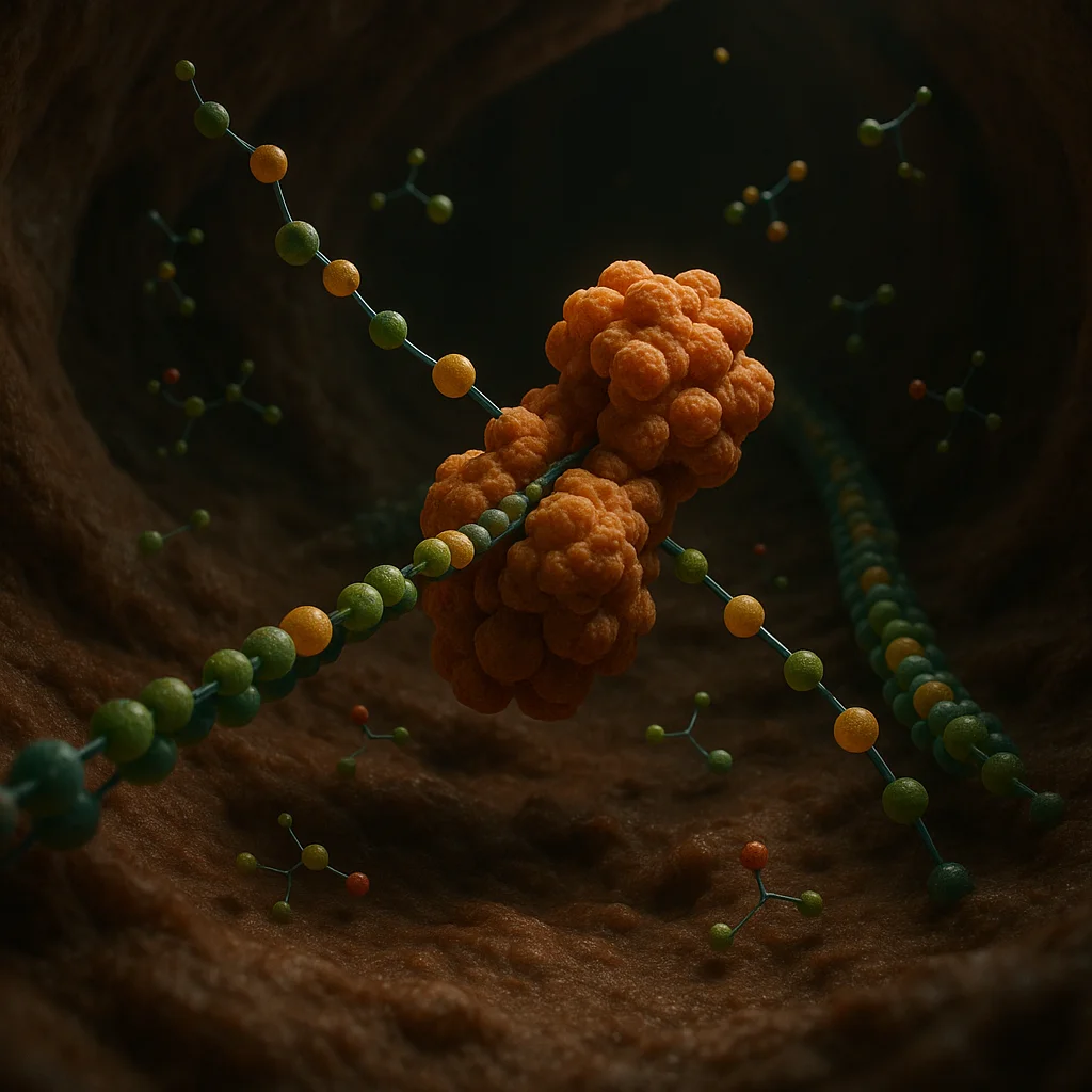
Edri et al. have meticulously traced folic acid’s intervention to the upregulation of the gene ALDH1L1, encoding the enzyme aldehyde dehydrogenase 1 like 1. This enzyme presides over a pivotal transformation. Specifically, ALDH1L1 enables the oxidation of retinaldehyde—a vitamin A (retinol) metabolite—into retinoic acid. Chemically, this can be represented as:
Retinaldehyde + NAD⁺ + H₂O → Retinoic acid + NADH + 2H⁺
Retinoic acid exerts regulatory command over gene expression through nuclear retinoic acid receptors (RARs), orchestrating spatial and temporal patterns of cellular proliferation, differentiation, and migration in the neural ectoderm.
The researchers employed *Xenopus laevis* embryos, a robust in vivo model for vertebrate neural development, to interrogate this pathway. Embryos were engineered to exhibit neural tube closure failures. Exogenous folic acid supplementation rectified the closure in a significant subset. However, upon CRISPR-mediated disruption of ALDH1L1, this phenotypic rescue was abrogated—demonstrating that ALDH1L1 activity is requisite for the protective function of folic acid. The restoration of normalcy was observed only when both folic acid and ALDH1L1 were present, implying an obligate epistatic relationship.
Extending these observations, the team demonstrated that recombinant human ALDH1L1 could catalyze retinoic acid synthesis in vitro. Parallel experiments in mammalian cell lines revealed activation of the same pathway, further consolidating the relevance of these findings for human embryogenesis.
Disarray in this axis triaged dramatic alterations at the cellular level. Deficient retinoic acid signaling—induced via genetic or pharmacological intervention—provoked unchecked proliferation in the neural plate, producing hyperplasia and spatial aberrancy. The application of folic acid could normalize these perturbations, conditional on functional ALDH1L1; absent the enzyme, folic acid’s efficacy vanished.
Given ALDH1L1’s dependence on a vitamin A-derived substrate, the investigators conjectured synergism between folic acid and retinol supplementation. Low-dose retinol administered alongside suboptimal folic acid resulted in improved morphogenic outcomes in the embryonic models. Yet, caution resounds: vitamin A exhibits potent teratogenicity in excess, with developmental homeostasis contingent on stringent retinoic acid titration.
These breakthroughs reframe the dogma that folic acid functions merely as a metabolic supplement. The evidence delineates folic acid as integral to a tightly regulated enzyme-mediated induction of developmental signals, namely retinoic acid, at the neural tube closure juncture.
This mechanistic elucidation also gestures toward an explanation for the clinical observation that folic acid prophylaxis, while robust, is not universally preventive of neural tube defects. Intrinsic dysfunctions within the ALDH1L1–retinoic acid biosynthetic axis—be they genetic, epigenetic, or substrate-limited—may underlie residual susceptibilities in a subset of pregnancies. Establishing the prevalence and penetrance of such defects in the human population, and their amenability to therapeutic modulation, remains an imperative trajectory for future research.
Thus, Edri and colleagues’ findings fundamentally advance the molecular understanding of folic acid’s protective role in embryonic neurodevelopment. By uncovering its reliance on ALDH1L1-mediated retinoic acid synthesis, this work not only unlocks new investigative avenues but also reframes the strategic landscape of prenatal nutritional policy and intervention.
The foundational problem arises in one of embryogenesis’s most sensitive events: the morphogenesis of the neural tube, scaffolding for the future central nervous system. This process begins with the neural plate, a monolayer of precursor cells, folding dorsally. The “zippering” closure of its edges is precarious; disruptions yield catastrophic consequences, typified by anencephaly or spina bifida. Here, folic acid has long demonstrated a protective effect, though its mode of action eluded definitive mechanistic description.
Edri et al. have meticulously traced folic acid’s intervention to the upregulation of the gene ALDH1L1, encoding the enzyme aldehyde dehydrogenase 1 like 1. This enzyme presides over a pivotal transformation. Specifically, ALDH1L1 enables the oxidation of retinaldehyde—a vitamin A (retinol) metabolite—into retinoic acid. Chemically, this can be represented as:
Retinaldehyde + NAD⁺ + H₂O → Retinoic acid + NADH + 2H⁺
Retinoic acid exerts regulatory command over gene expression through nuclear retinoic acid receptors (RARs), orchestrating spatial and temporal patterns of cellular proliferation, differentiation, and migration in the neural ectoderm.
The researchers employed *Xenopus laevis* embryos, a robust in vivo model for vertebrate neural development, to interrogate this pathway. Embryos were engineered to exhibit neural tube closure failures. Exogenous folic acid supplementation rectified the closure in a significant subset. However, upon CRISPR-mediated disruption of ALDH1L1, this phenotypic rescue was abrogated—demonstrating that ALDH1L1 activity is requisite for the protective function of folic acid. The restoration of normalcy was observed only when both folic acid and ALDH1L1 were present, implying an obligate epistatic relationship.
Extending these observations, the team demonstrated that recombinant human ALDH1L1 could catalyze retinoic acid synthesis in vitro. Parallel experiments in mammalian cell lines revealed activation of the same pathway, further consolidating the relevance of these findings for human embryogenesis.
Disarray in this axis triaged dramatic alterations at the cellular level. Deficient retinoic acid signaling—induced via genetic or pharmacological intervention—provoked unchecked proliferation in the neural plate, producing hyperplasia and spatial aberrancy. The application of folic acid could normalize these perturbations, conditional on functional ALDH1L1; absent the enzyme, folic acid’s efficacy vanished.
Given ALDH1L1’s dependence on a vitamin A-derived substrate, the investigators conjectured synergism between folic acid and retinol supplementation. Low-dose retinol administered alongside suboptimal folic acid resulted in improved morphogenic outcomes in the embryonic models. Yet, caution resounds: vitamin A exhibits potent teratogenicity in excess, with developmental homeostasis contingent on stringent retinoic acid titration.
These breakthroughs reframe the dogma that folic acid functions merely as a metabolic supplement. The evidence delineates folic acid as integral to a tightly regulated enzyme-mediated induction of developmental signals, namely retinoic acid, at the neural tube closure juncture.
This mechanistic elucidation also gestures toward an explanation for the clinical observation that folic acid prophylaxis, while robust, is not universally preventive of neural tube defects. Intrinsic dysfunctions within the ALDH1L1–retinoic acid biosynthetic axis—be they genetic, epigenetic, or substrate-limited—may underlie residual susceptibilities in a subset of pregnancies. Establishing the prevalence and penetrance of such defects in the human population, and their amenability to therapeutic modulation, remains an imperative trajectory for future research.
Thus, Edri and colleagues’ findings fundamentally advance the molecular understanding of folic acid’s protective role in embryonic neurodevelopment. By uncovering its reliance on ALDH1L1-mediated retinoic acid synthesis, this work not only unlocks new investigative avenues but also reframes the strategic landscape of prenatal nutritional policy and intervention.