Field: Technology
A revolutionary new microscope could reveal the hidden machinery of life
Published | Technical Staff
Visualization
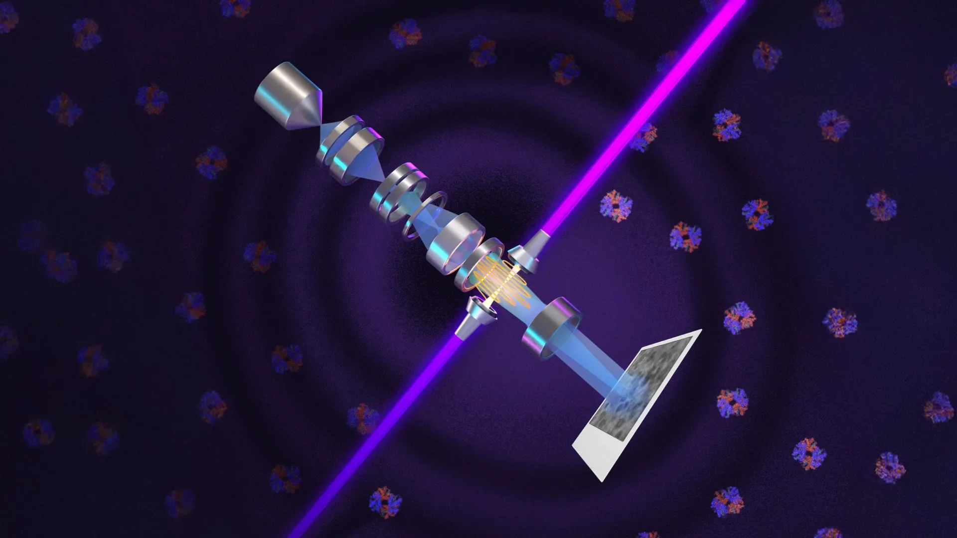
In a groundbreaking advancement poised to transform our understanding of cellular biology, researchers at the University of California, Berkeley have engineered a laser-powered microscope that promises to unveil intricate structures within human cells, which have eluded scientists for decades. This innovative apparatus, known as Theia, harnesses the principles of laser phase plates to drastically enhance the capabilities of cryoelectron microscopy (cryo-EM) and cryoelectron tomography (cryo-ET). By significantly increasing image clarity and contrast, it allows for detailed imaging of the minuscule proteins that conventional electron microscopy often struggles to capture.
Phase contrast microscopy, a technique crucial for resolving faint cellular features, laid the groundwork for this leap forward nearly a century ago. Developed by Dutch physicist Frits Zernike, phase contrast relies on detecting phase shifts—minute delays that light experiences when traversing biological materials—thereby transforming transparency into discernible contrast. The resulting technique revolutionized biological imaging, earning Zernike a Nobel Prize in 1953. Yet, when physicists attempted to apply similar phase-shifting methodologies to electron microscopy, they faced significant hurdles. The first attempts suffered from instability, compromised resolution, and weakened electron beams that failed to provide sufficient clarity.
Müller and his colleague Robert Glaeser began to sketch a solution over a decade ago that would leverage high-intensity lasers not merely for illumination, but as agents to manipulate the phase of the electrons in the beam. The theoretical underpinning revolves around the interaction between the focused laser and the electron beam itself, resulting in refined contrast that could enable biologists to unequivocally identify and differentiate small proteins and cellular components that were previously indistinguishable.
The heart of this advancement lies in what the researchers label as the world's most intense focused continuous-wave laser, which interacts with the microscope's electron beam to induce a phase alteration. This improvement effectively enhances the signal-to-noise ratio, particularly beneficial for examining the crowded interiors of cells, which might be likened to navigating a complex forest in search of individual leaves amidst the verdant chaos. As cryo-ET requires a leap in imaging contrast to elucidate the three-dimensional organization of molecular structures, this laser phase plate serves as a beacon of promise, transitioning low-contrast images into stark revelations of biological machinery.
With financial backing from Biohub, Müller equipped his cryo-EM apparatus with this advanced laser phase plate. Not far away, Bridget Carragher is orchestrating the creation of a complementary microscope design at Biohub's imaging facilities, employing a dual-laser configuration aimed at further enhancing contrast while safeguarding optical component integrity. Each laser operates at approximately half the power yet is orientated perpendicularly, offering a symbiotic partnership that mitigates potential distortions and thermal wear.
The implications of Theia mark an epochal step forward in biological imaging. Müller characterizes his invention as the “Formula 1 microscope” due to its advanced electron optics, which already furnish superior resolution relative to traditional cryo-EM frameworks. The anticipated synergy of its laser phase plate heralds the potential for this microscope to become an unparalleled instrument for elucidating the complexities of life at a molecular level. Recent technical findings and pioneering images produced by this avant-garde apparatus have been highlighted in the journal Science, while the intricacies of Biohub's twin-laser instrument are available in a contemporaneously posted research preprint.
These advancements come as scientific inquiry grapples with a fundamental limitation intrinsic to biological imaging: cells' high water content confers transparency, thereby complicating the visualization of crucial subcellular structures. Historical strategies often involved staining cells to enhance contrast, a practice that invariably leads to cellular death and compromises the fidelity of observed dynamics. The introduction of a laser-driven phase-contrast framework within the electron microscopy domain addresses these challenges while retaining the viability of individual cellular components.
As researchers embark on a new era of micro-imaging, the convergence of theoretical innovation and technological prowess offers a promising frontier. With the capacity to inspect proteins on the scale of one-third of previously captured sizes, the applications of the laser phase plate promise to illuminate the interactions and arrangements of biomolecules within their native cellular environments. This intricate dance of molecular machinery, revealed through enhanced imaging techniques, could catalyze significant advancements in drug discovery and our comprehension of the intricate tapestry of life itself. Theia, and its contemporaneous counterparts, open unprecedented windows into the microscopic worlds that constitute life, propelling the scientific community into exhilarating new realms of discovery.
Phase contrast microscopy, a technique crucial for resolving faint cellular features, laid the groundwork for this leap forward nearly a century ago. Developed by Dutch physicist Frits Zernike, phase contrast relies on detecting phase shifts—minute delays that light experiences when traversing biological materials—thereby transforming transparency into discernible contrast. The resulting technique revolutionized biological imaging, earning Zernike a Nobel Prize in 1953. Yet, when physicists attempted to apply similar phase-shifting methodologies to electron microscopy, they faced significant hurdles. The first attempts suffered from instability, compromised resolution, and weakened electron beams that failed to provide sufficient clarity.
Müller and his colleague Robert Glaeser began to sketch a solution over a decade ago that would leverage high-intensity lasers not merely for illumination, but as agents to manipulate the phase of the electrons in the beam. The theoretical underpinning revolves around the interaction between the focused laser and the electron beam itself, resulting in refined contrast that could enable biologists to unequivocally identify and differentiate small proteins and cellular components that were previously indistinguishable.
The heart of this advancement lies in what the researchers label as the world's most intense focused continuous-wave laser, which interacts with the microscope's electron beam to induce a phase alteration. This improvement effectively enhances the signal-to-noise ratio, particularly beneficial for examining the crowded interiors of cells, which might be likened to navigating a complex forest in search of individual leaves amidst the verdant chaos. As cryo-ET requires a leap in imaging contrast to elucidate the three-dimensional organization of molecular structures, this laser phase plate serves as a beacon of promise, transitioning low-contrast images into stark revelations of biological machinery.
With financial backing from Biohub, Müller equipped his cryo-EM apparatus with this advanced laser phase plate. Not far away, Bridget Carragher is orchestrating the creation of a complementary microscope design at Biohub's imaging facilities, employing a dual-laser configuration aimed at further enhancing contrast while safeguarding optical component integrity. Each laser operates at approximately half the power yet is orientated perpendicularly, offering a symbiotic partnership that mitigates potential distortions and thermal wear.
The implications of Theia mark an epochal step forward in biological imaging. Müller characterizes his invention as the “Formula 1 microscope” due to its advanced electron optics, which already furnish superior resolution relative to traditional cryo-EM frameworks. The anticipated synergy of its laser phase plate heralds the potential for this microscope to become an unparalleled instrument for elucidating the complexities of life at a molecular level. Recent technical findings and pioneering images produced by this avant-garde apparatus have been highlighted in the journal Science, while the intricacies of Biohub's twin-laser instrument are available in a contemporaneously posted research preprint.
These advancements come as scientific inquiry grapples with a fundamental limitation intrinsic to biological imaging: cells' high water content confers transparency, thereby complicating the visualization of crucial subcellular structures. Historical strategies often involved staining cells to enhance contrast, a practice that invariably leads to cellular death and compromises the fidelity of observed dynamics. The introduction of a laser-driven phase-contrast framework within the electron microscopy domain addresses these challenges while retaining the viability of individual cellular components.
As researchers embark on a new era of micro-imaging, the convergence of theoretical innovation and technological prowess offers a promising frontier. With the capacity to inspect proteins on the scale of one-third of previously captured sizes, the applications of the laser phase plate promise to illuminate the interactions and arrangements of biomolecules within their native cellular environments. This intricate dance of molecular machinery, revealed through enhanced imaging techniques, could catalyze significant advancements in drug discovery and our comprehension of the intricate tapestry of life itself. Theia, and its contemporaneous counterparts, open unprecedented windows into the microscopic worlds that constitute life, propelling the scientific community into exhilarating new realms of discovery.